What it is
The optic nerve is the cable that carries images from the eye to the brain. It is made up of more than a million fibres. In glaucoma those fibres die off little by little, and with them you lose patches of your field of vision.
This guide covers the most common type, primary open-angle glaucoma. It nearly always affects both eyes, although one may be further along than the other. It is usually linked to a pressure inside the eye that is higher than the nerve can tolerate. But not always: some people with normal pressure develop it, and some people with high pressure never do.
If you have been told your pressure is high but the nerve is healthy, that is called ocular hypertension, and it has its own guide: ocular hypertension and glaucoma suspects.
Why it gives no warning
Glaucoma starts with your side vision, the peripheral vision. The centre, the part you use to read or watch TV, holds out until the end. On top of that, each eye covers the gaps in the other and the brain fills in what is missing. The result: you notice nothing for years.
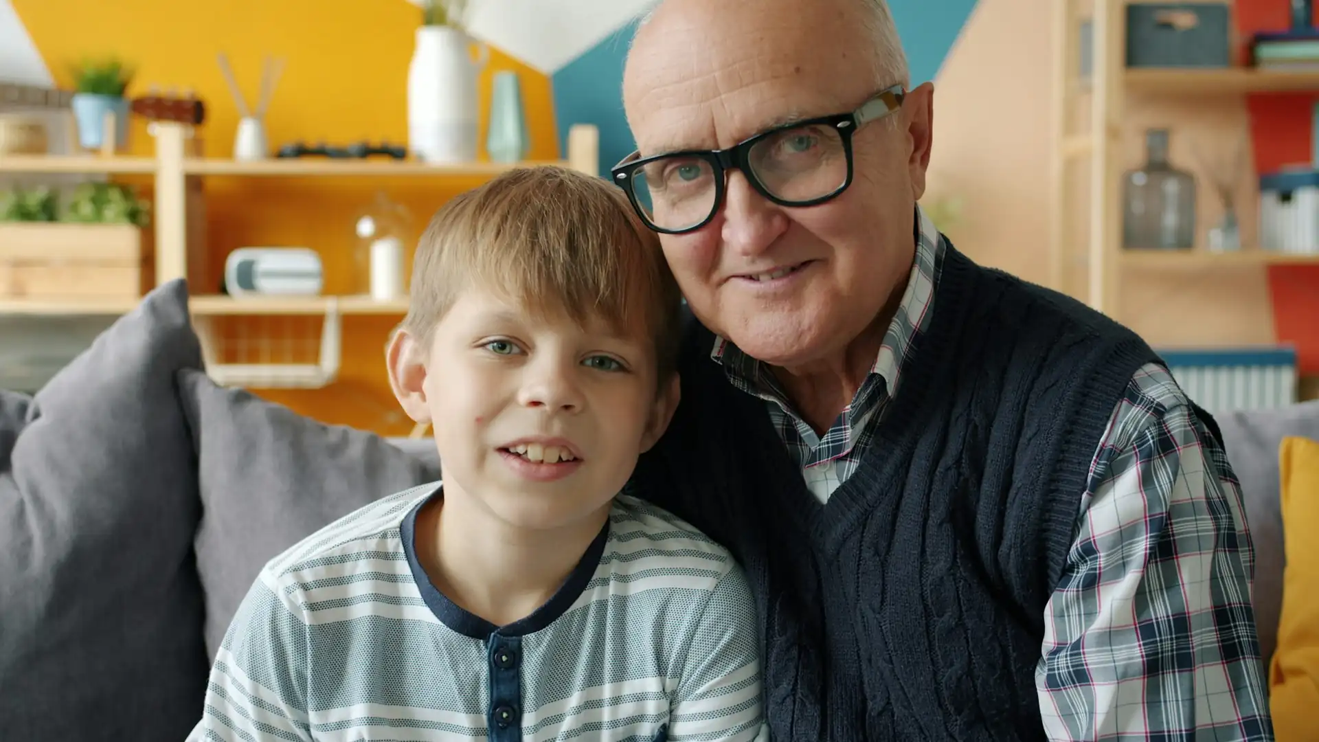 Normal vision
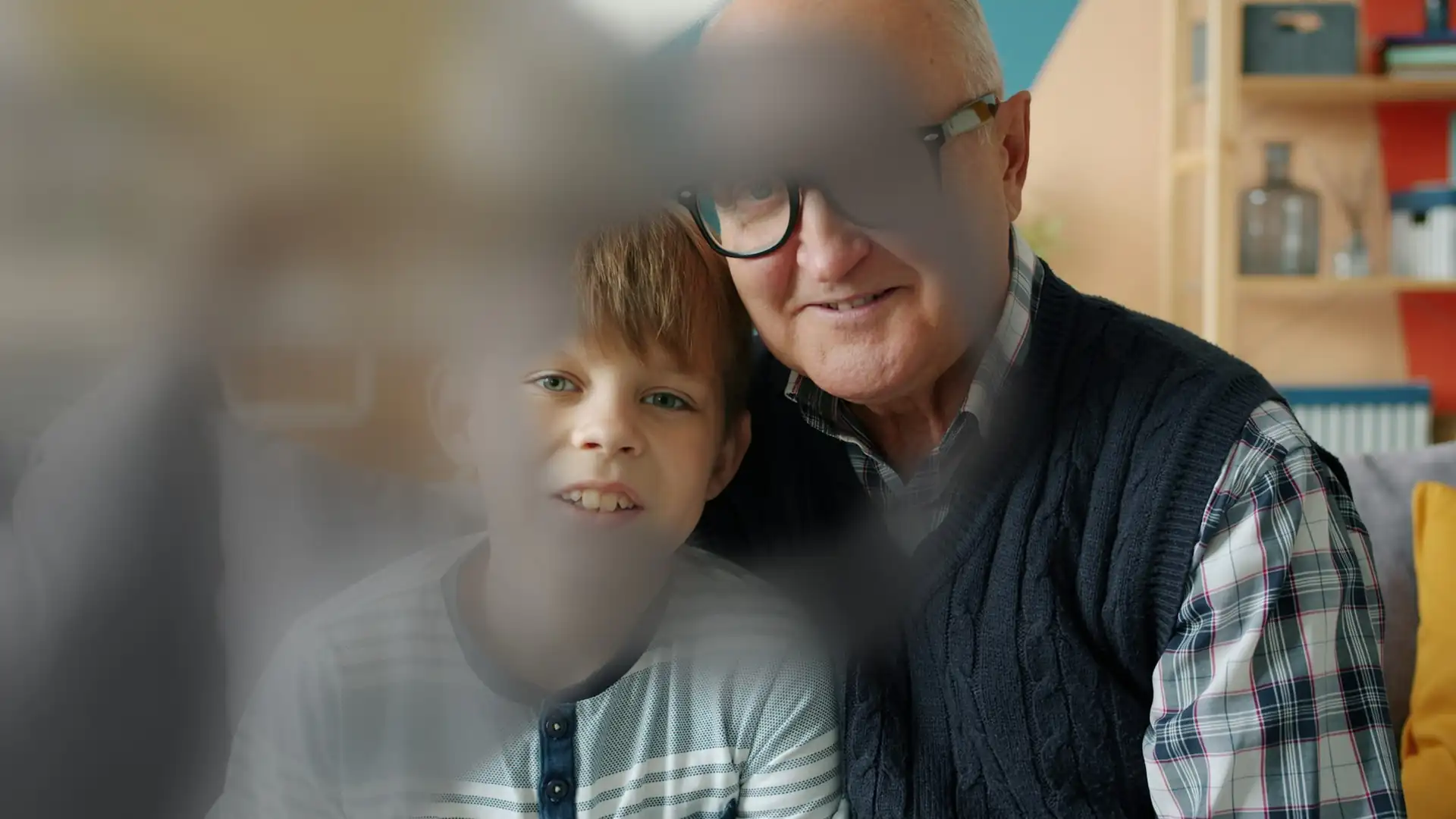 With glaucoma
It does not hurt. The eye does not go red. Near and distance vision stay the same. By the time someone realises they are bumping into things or missing a car coming from the side, the nerve has already lost a good part of its fibres.
The only way to catch it in time is to have your eyes checked. From the age of 40, and earlier if you have relatives with glaucoma, it is worth having the eye pressure measured and the optic nerve examined even if you see well.
Risk factors
- High eye pressure. It is the most important factor and the only one that can be treated. The higher it is, the greater the risk.
- Age. The risk clearly goes up over the years.
- Having parents or siblings with glaucoma. In a study that examined the brothers and sisters of glaucoma patients, having it in the family made it about nine times more likely. If that is you, tell your ophthalmologist and get checked even if you notice nothing.
- Ethnic background. It is considerably more common, and appears earlier, in people of African or Latin American descent.
- Short-sightedness (myopia), especially if it is high.
- Diabetes and low blood pressure. With low blood pressure the optic nerve gets less blood flow.
- A thin or less elastic cornea. The cornea is the clear window at the front of the eye, and its thickness and behaviour affect both the risk and how the pressure is measured.
Having one of these factors does not mean you will get glaucoma. It means it is worth keeping an eye on.
How it is diagnosed
There is no single test. The diagnosis comes from putting several pieces together, and none of them hurt:
- The eye pressure (intraocular pressure). With ORA technology I measure it taking the stiffness of the cornea into account, which gives a more reliable reading than the standard measurement.
- The thickness and biomechanics of the cornea. A thin cornea can make the pressure look lower than it really is. The ORA also measures how well the cornea cushions (known as hysteresis), and a low value is linked to higher risk.
- The optic nerve, looking at it directly at the back of the eye.
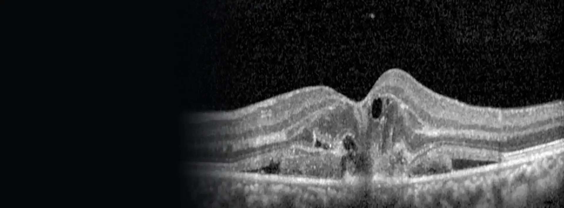
- OCT, which measures in microns the thickness of the nerve fibre layer around the nerve and in the macula. It can pick up fibre loss before any gaps appear in the visual field.
- Perimetry, or the visual field test. You look at a fixed point and press a button every time you see a light appear at the sides. This draws a map of what each eye can see.
- Gonioscopy, a lens placed on the eye to look at the angle where the fluid drains out. It tells open-angle glaucoma apart from closed-angle glaucoma, which is treated differently.
The first visit gives us a starting point. What really confirms or rules out damage that is progressing is comparing the tests over time.
Treatment
Every treatment aims for the same thing: to bring the eye pressure down to a level at which the nerve stops being damaged. We call that level the target pressure, and it is different for each person depending on the state of the nerve and their risk factors. A common starting point is to lower it by 20 to 30% from where it began, then adjust depending on how things go.
This applies to normal-tension glaucoma too: even when the number already looks fine, lowering it further protects the nerve.
Eye drops
This is the usual way to start. There are several groups of eye drops and many are used just once a day. If one is not enough or does not suit you, it is changed or combined with another.
Their big weakness is that you have to use them every day, for years, and glaucoma does not remind you that you have it. Many patients miss doses without realising. If you find it hard, if they sting or if they run out before they should, tell me: it can almost always be sorted out.
Laser: selective laser trabeculoplasty (SLT)
This is a gentle laser applied to the area where fluid drains out of the eye so that it flows out better. It can be used as the first treatment, instead of drops, or to reduce the number of drops you need. Its effect can wear off over the years, and it can be repeated.
If the angle of the eye is narrow
There is another type of glaucoma in which the problem is that the fluid's drainage route is very closed. To open it I carry out, in the clinic and with a laser, an iridotomy (a tiny opening in the iris so that fluid can circulate) or an iridoplasty (which pulls back the edge of the iris to clear the angle). They are quick procedures, done with anaesthetic drops. I explain them in detail in the guide on narrow angles and angle closure.
Surgery
When drops and laser are not enough to stop the damage, there are several operations that create a new drainage route for the fluid or fit small drainage devices.
What about supplements or natural remedies? At present there is no solid evidence that they help. They are not a substitute for treatment.
Putting your drops in properly
One drop put in properly is worth more than three put in badly. Getting it right is not as easy as it looks, especially as you get older or if your hands are shaky.
How to put in your eye drops
- Wash your hands.
- Tilt your head back, or lie down.
- Gently pull down your lower eyelid to make a small pocket.
- Let one drop fall into that pocket without the bottle touching your eye or lashes.
- Close your eye gently, without squeezing, and press with one finger on the tear duct (the corner next to your nose) for a couple of minutes. This keeps the drop in the eye and means less of it gets into your bloodstream.
- If you use two different eye drops, wait about 5 minutes between them.
Always put them in at the same time, tied to something you do every day (breakfast, brushing your teeth). An alarm on your phone helps. And bring the bottles to your check-up.
Check-ups
Glaucoma is controlled, not cured. Check-ups are there to make sure the pressure stays where it should and the nerve is not losing more fibres. At every visit I check the pressure and, as often as needed, I repeat the OCT and the visual field test.
How often depends on you. If the pressure is at its target and the nerve has not changed for more than six months, check-ups are usually every 6 to 12 months. If the glaucoma is progressing, visits come closer, every 1 to 3 months until it is under control, and advanced glaucoma is watched more closely. At the start it is worth repeating the visual field test more often, because that shows sooner who is progressing quickly.
- Do not stop your drops even if you feel fine. Not noticing anything is exactly what we expect.
- If you are prescribed a new medicine, or have an operation on your eye for something else, let me know.
- Tell your parents, brothers and sisters, and children. They should have their eyes checked too.
Do you have relatives with glaucoma, or have you been told the pressure in your eye is high?
Call and I will give you an appointment. In the same visit I measure the eye pressure and examine the optic nerve.