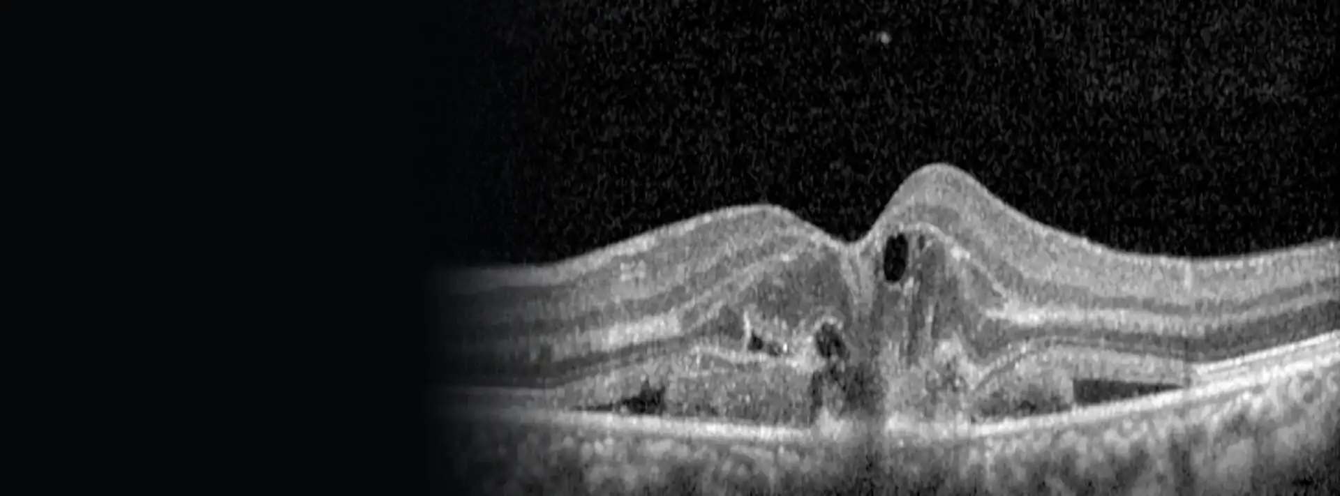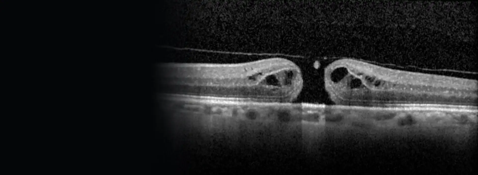
What it is
The macula is the centre of the retina, the part you use to read, thread a needle or recognise a face. A macular hole is a very small, round break right in its centre. It measures less than half a millimetre, but it is in the worst possible place.
It affects your central vision. Your peripheral vision, out to the sides, is kept, so it does not make you blind: you can still get around at home and outside.
Not everything called a hole is one
Two conditions look similar but are different. A lamellar hole is a defect that does not go all the way through the retina. A pseudohole is a false appearance of a hole created by an epiretinal membrane pulling on the edges. Neither behaves like a full hole, and the OCT tells them apart without any doubt.
Why it happens
The eye is filled with a gel, the vitreous, which comes away from the retina as we get older. Normally this happens without any trouble. Sometimes it separates around the macula but stays stuck in the centre, and that point of attachment pulls until the retina opens.
It usually appears after the age of 55 and is quite a bit more common in women. It is almost always "idiopathic", meaning there is no other cause behind it. Less often it follows a blow to the eye, and then it tends to affect younger people.
It forms in stages, over weeks or months:
- Impending hole: the vitreous pulls and the retina is distorted, but it has not broken yet. About half of these cases sort themselves out when the vitreous lets go.
- Small hole: there is now a full break, though smaller than 0.4 mm. Most grow if they are not treated.
- Large hole: wider, often with fluid around it. Vision drops further.
Symptoms
It is usually noticed in one eye only, and sometimes quite suddenly:
- Lines and letters look distorted or bent in the centre.
- A blurred or grey spot right where you fix your gaze.
- Blurred central vision, which makes reading or recognising faces difficult.
- Things look smaller or larger with that eye.
Because the other eye makes up for it, some people only find out when they happen to cover one eye. That is why it is worth checking each eye separately from time to time, for example with the Amsler grid.
If a new spot or distortion appears in the centre of your vision, book an appointment soon. It is not a matter of hours, but it is not something to leave either: a recent, small hole closes better and leaves more vision than one that has been there for many months. This also applies to the other eye if you have already had a hole.
How it is diagnosed

I examine the back of the eye with the pupil dilated and do an OCT scan. This is the deciding test: in seconds, and without touching the eye, it confirms whether there is a full hole, measures its size and shows whether the vitreous is still pulling or whether there is a membrane. With that information I choose the treatment, and I can show it to you on the screen.
I always check the other eye too. Between 1 and 2 in 10 people with a hole go on to develop one in the other eye in the following years. The risk is lower if the vitreous in that eye has already fully come away, and the OCT helps me find out.
What happens without treatment
A full hole almost never closes on its own; it happens in a few cases, mainly when it is very small. What usually happens is that it grows and the vision in that eye drops to the point where you cannot read the large letters on the chart, and then it settles there. Your side vision is kept.
With an impending hole, on the other hand, the right approach is to keep an eye on it with check-ups every few months. Operating at that stage has not been shown to stop it from opening, and in many cases it resolves on its own.
There are no drops or tablets that can replace surgery. Closure with anti-inflammatory drops has been described in some very small holes with swelling, but these are isolated cases.
The operation
When
When there is a full hole, the general recommendation is to operate. It does not have to be done the same day, but it is not a good idea to wait many months either: the longer it has been open, the less well it closes and the less vision comes back.
How
The operation is a vitrectomy. Using very fine instruments, 23G, 25G or 27G, I enter the eye through three tiny openings and do three things:
- I remove the vitreous and release the pull on the macula.
- I peel the internal limiting membrane, a clear layer on the surface of the retina. I use a dye to see it. Removing it makes closure more likely and lowers the chance of the hole opening again.
- At the end I fill the eye with a gas bubble, which gently presses the edges of the hole together while it heals.
For large holes, or ones that have already been operated on without success, there are extra techniques, such as using a small flap of that same membrane to cover the hole.
It is done under local anaesthesia with a retrobulbar injection, behind the eye, which numbs it completely. You can watch a real operation in the vitrectomy with ILM peeling video.
If you also have a cataract
Almost everyone who still has their natural lens develops a cataract in the years after a vitrectomy. That is why it is sometimes worth operating on the cataract at the same time; studies show no difference in results compared with doing them separately. We will weigh this up together.
Posture and the gas bubble
Posture
The bubble floats. For it to press on the macula, your head needs to be in the right position for the first few days. People used to be asked to stay face down for two weeks. We now know that for small holes much less is enough, and that what matters is not lying on your back. For large holes, keeping your face down for a few days does seem to help.
I will tell you which position to keep and for how long, depending on the size of your hole. I explain this before we schedule the operation, so you can make arrangements at home.
While there is gas in your eye
Your vision in that eye will be very blurred. Depending on the gas, the bubble takes from a few days to several weeks to be absorbed, and your vision clears as it gets smaller. While it lasts:
- Do not fly or go up into the mountains or over high passes: the gas expands with altitude and the pressure in the eye can rise dangerously.
- If you need an operation for something else under general anaesthesia, tell them you have gas in your eye. There is one anaesthetic (nitrous oxide) that cannot be used.
- Do not drive until I tell you that you can.
Results and risks
With modern surgery more than 9 in 10 holes close. Once closed, most patients recover a good deal of vision, often enough to read, and the improvement can continue for months, even years. Even so, vision is not usually perfect: some blurring or distortion may remain.
Case 1
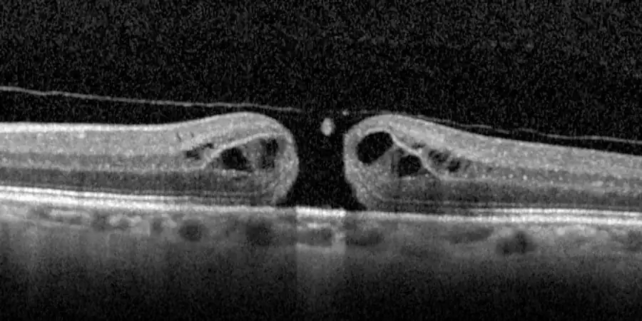 Before
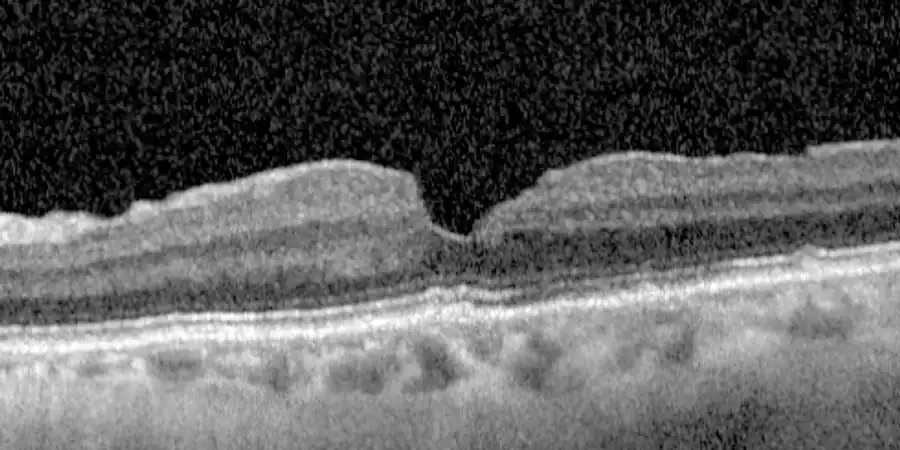 After
AfterCase 2
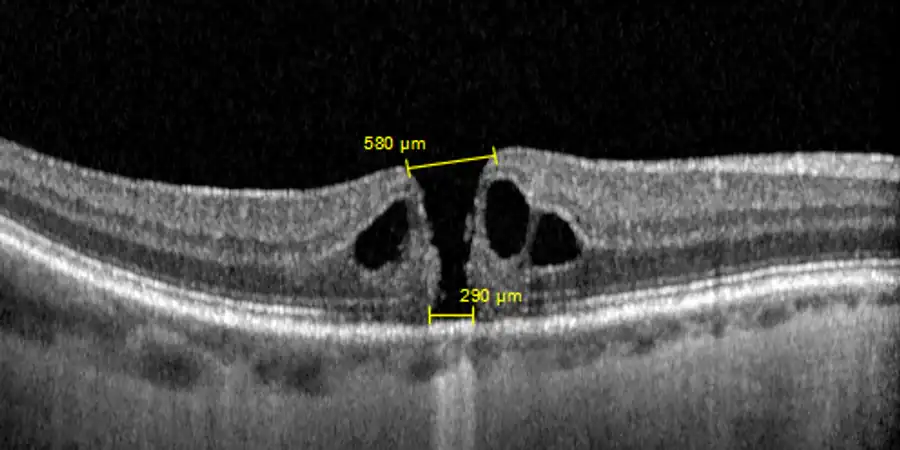 Before
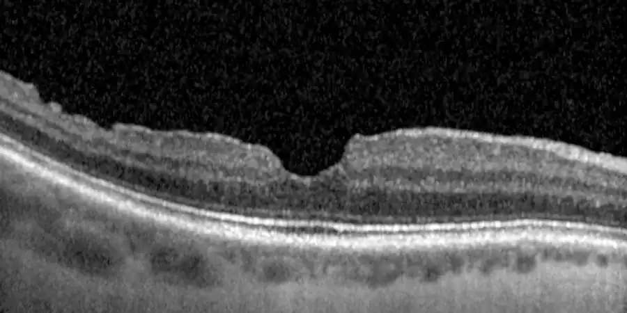 After
AfterIf the hole does not close the first time, a second operation succeeds in many cases, although the vision gained is usually less.
The risks we discuss before operating:
- Cataract: the most common consequence in people who still have their natural lens.
- The hole opening again: up to 1 in 10, fewer when the internal limiting membrane is peeled.
- Retinal tear or detachment: uncommon, and almost always repaired without the hole reopening.
- High pressure in the eye in the first few days, especially with gas and in people with glaucoma.
- Serious infection inside the eye (endophthalmitis): very rare, fewer than 1 in 2000 operations.
I see you the day after the operation, again after a week or two, and then depending on how things go, always with an OCT to check the hole has closed.
Call straight away if, after the operation, you have pain that is getting worse, your vision gets worse instead of clearing, or you notice lots of new floaters or a shadow to one side. These can be signs of high pressure, infection or retinal detachment.
Do you notice a spot, or letters looking bent, in the centre of your vision?
Call and I will give you an appointment. With an OCT scan at the same visit I can tell whether there is a hole, how big it is and what is best to do.